Aortic injuries following road traffic accidents are rare but extremely serious. In many cases, these injuries can become life-threatening within a short time if they are not identified and treated quickly.
Recently, a 42-year-old gentleman presented with an alleged history of a road traffic accident. On evaluation, he was diagnosed with blunt chest trauma, which had caused injuries to the descending thoracic aorta, along with rib fractures and a left-sided hemothorax, which means blood had collected in the chest cavity.
The patient also had associated splenic and renal injuries. A general surgery opinion was sought, and conservative management was planned for those injuries.
CT Aortogram Revealed Serious Aortic Injury
To further assess the aortic injury, a CT aortogram was performed. The scan revealed two separate intimal tears in the descending thoracic aorta, located approximately 3 cm and 6 cm from the left subclavian artery.
More importantly, the second tear was causing a near-total occlusion of the true lumen of the aorta. This had started affecting blood supply to vital areas, leading to signs of renal and lower limb malperfusion.
In simple terms, the injury was not only affecting the aorta but was also reducing blood flow to the kidneys and lower limbs, making the situation highly critical.
Timely Decision to Perform TEVAR
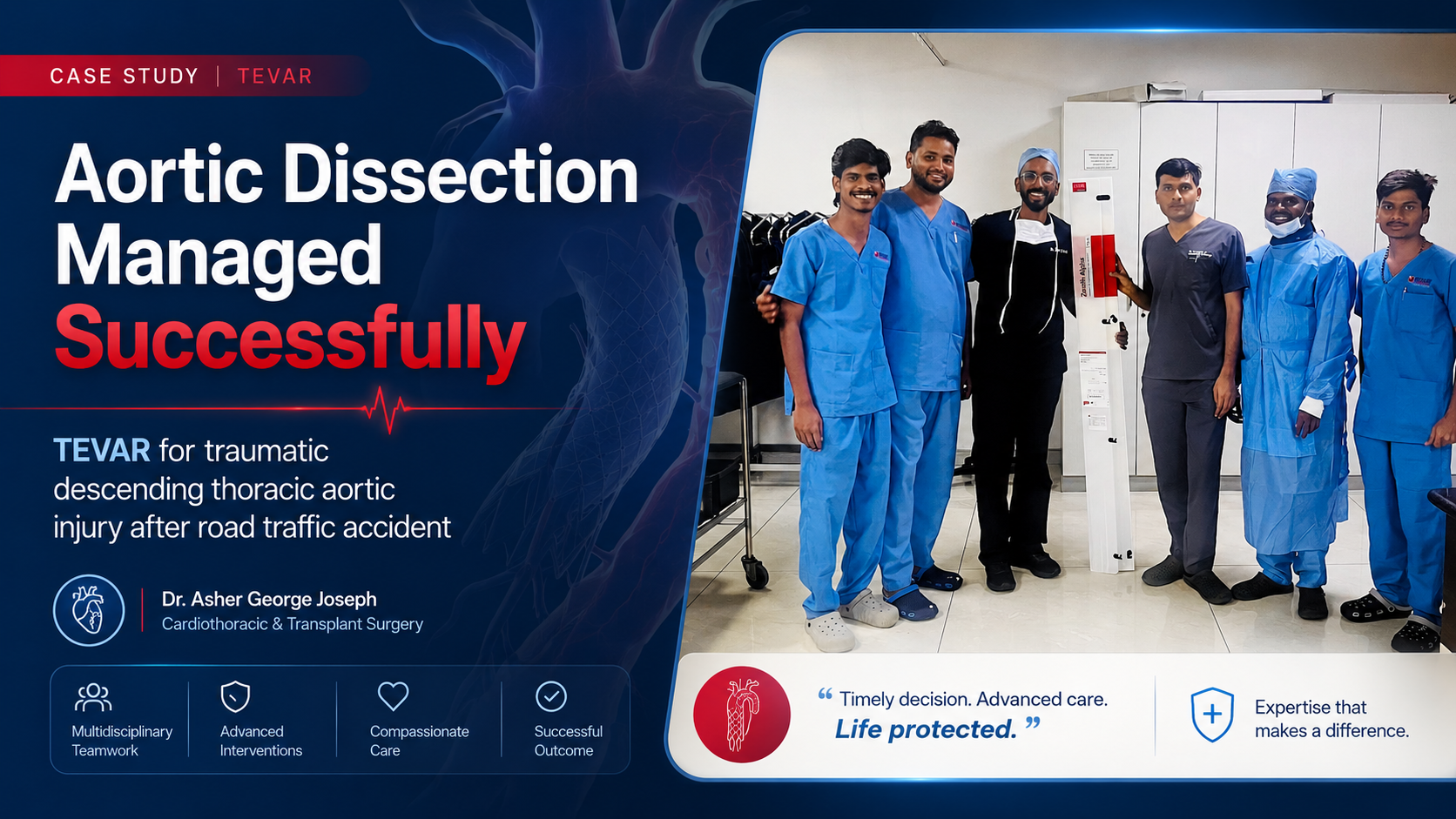
After detailed discussions between the cardiothoracic surgery team and Dr. Naveen, Interventional Radiology Consultant, the decision was taken to treat the Type B aortic dissection using TEVAR.
TEVAR, or Thoracic Endovascular Aortic Repair, is a minimally invasive procedure used to treat certain diseases and injuries of the thoracic aorta. Instead of performing a large open surgery, a stent graft is placed inside the aorta through the blood vessels to seal the tear and restore proper blood flow.
The patient and family were counselled regarding the critical nature of the condition, the grave prognosis, and the planned procedure. After obtaining informed consent, the procedure was performed under general anaesthesia.
Successful Stent Deployment and Recovery
TEVAR was performed using a single stent graft. After successful stent deployment, the left hemothorax was drained using an intercostal drain.
The patient tolerated the procedure well. Most importantly, his renal function and distal limb blood flow improved immediately after the stent placement.
He was closely observed in the ICU for 48 hours. After the drain was removed and his condition remained stable, he was shifted to the ward for further recovery.
The patient continued to improve and was successfully discharged on postoperative day 4.
Why This Case Is Important
This case highlights the importance of early diagnosis, rapid decision-making, and a coordinated team approach in managing life-threatening aortic injuries.
A traumatic aortic dissection can progress quickly and may compromise blood flow to vital organs. In such situations, timely intervention can make a major difference in patient outcomes.
This result was possible because of effective collaboration between the cardiothoracic surgery, interventional radiology, critical care, and anaesthesia teams.
Teamwork in complex emergencies directly translates to timely decision-making, effective patient care, and better recovery.